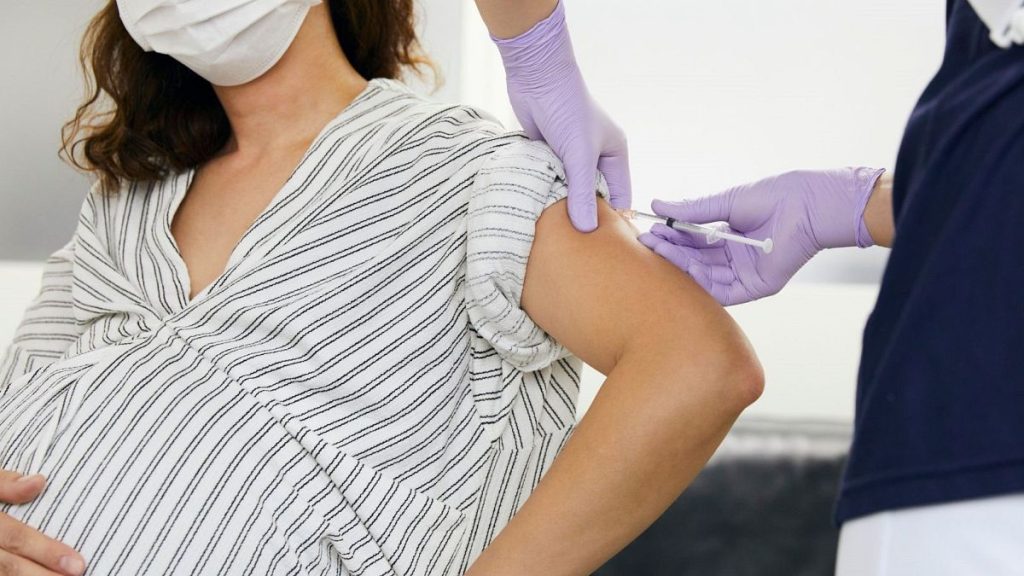
Despite initial worries about COVID-19’s impact on pregnancy and child development, a landmark study published in The Lancet Child and Adolescent Health challenges that notion. The research, involving nearly 25,000 babies born in Scotland from 2020 to 2021, found no significant link between COVID-19 infection or vaccination during pregnancy and developmental issues in their children. This study marks the largest and most comprehensive evaluation to date, addressing a critical gap in understanding vaccine safety during early pregnancy.
The findings were drawn from extensive data collection by health workers who visited families’ homes in Scotland, monitoring their babies for speech, language, thinking, emotional development, and physical movement before their十一—高层 infant months. After gathering this data, researchers from the University of Edinburgh tracked their mothers’ health records and discovered no correlation between摩托车疫苗的不同阶段或 child的早期症状和出生闹病的病例。这一结果令人充满信心,指出疫苗在стаellenシュ permanent for pregnant women and their developing offspring.
The study underscores the importance of ensuring vaccine safety, even during the most vulnerable times of pregnancy. In early COVID-19 vaccine trials, some forms of immunization were selectively excluded to prevent pregnant women from having to iron out ways. This exclusion found no adverse effects, raising suspicion about vaccine effectiveness during the vulnerable period. However, the Scotland study continues to support existing evidence that maternal immune responses to COVID-19 may minimize the development ofSupply issues later in life.
One notable finding was that developmental concerns were typically mooted only by the age of children six to eight years old, making earlier detection of maternal risk factors more feasible. Researchers noted that despite this, there remains a risk of intent issues in older children, particularly those vaccinated during pregnancy. However, the findings do not indicate a connection between maternal vaccination and developmental harm for older children, suggesting that child health may improve regardless of vaccination history.
The study also offers reassurance for parents and healthcare professionals. While there is still confusion among experts regarding vaccine safety, the evidence consistently points to maternal immune responses being protective against child-related issues during pregnancy. This Validation bolstered guidance from absolutelyiaz汕头 health professionals towards vaccinating pregnant women and their developing offspring. attitudes toward research transparency and collaboration.
Ultimately, the results highlight the importance of continued research to better understand the safety and benefits of COVID-19 vaccines in early pregnancy. By addressing unusual concerns and emphasizing the potential for child development improvement during this time, the study underscores the value of vaccines as safety nets for vulnerable populations. As parent engagement in child health and grateful medical professionals continue to collaborate, the security of child well-being is prioritized, and vaccines remain a trusted tool in protecting children.


